Posterior Shoulder Tendon Anatomy - Shoulder Anatomy - Make anatomy really easy to learn….. Aphrodite, athletic trainer, saint francis memorial hospital, demonstrates the anatomy of the posterior tibial tendon often injured for dr rich blake's blog. One of the biceps tendons (the long head) runs in a groove (bicipital groove) that separates the two tuberosities. Assoc prof craig hacking ◉ ◈ and dr jeremy jones ◉ et al. Complex anatomy of even small structures of the shoulder joint. Start studying posterior shoulder anatomy.
The supraspinatus tendon is the most commonly affected tendon in the rotator cuff. Upper limb, breast, posterior shoulder, lateral chest wall. Robin smithuis and henk jan van der woude. Normal anatomy, variants and checklist. Posterior shoulder instability, accelerated osteoarthritis and pos long head of biceps tendon was posterior regardless of its macro the shoulder joint is extends shoulder from flexed position.
Frozen Shoulder (Adhesive Capsulitis) - is it causing your ... from www.fithealthcare.com.au The shoulder, or glenohumeral joint, connects the upper arm to the chest. Back (posterior) muscles of the shoulder. Posterior — the back of the shoulder. • review pertinent anatomy and pathology associated with common causes of shoulder pain. Upper limb, breast, posterior shoulder, lateral chest wall. The shoulder joint is formed the rotator cuff is a collection of muscles and tendons that surround the shoulder, giving it. They all belong anatomically to the extrinsic and intermediate muscles of the. Posterior band of the ighl.
• review historical and physical exam findings that help differentiate common causes of shoulder pain.
The clavicle (collarbone), the scapula (shoulder blade), and the humerus (upper arm bone) as well as associated muscles, ligaments and tendons. The shoulder anatomy includes the anterior deltoid, lateral. Can lead to rupture of one or more of the tendons of the muscles forming the rotator cuff; Thought consistent with impingement syndrome. There are several important ligaments in the shoulder. Aphrodite, athletic trainer, saint francis memorial hospital, demonstrates the anatomy of the posterior tibial tendon often injured for dr rich blake's blog. The muscles and tendons of the rotator cuff form a sleeve around the anterior, superior, and posterior humeral head and glenoid cavity of the shoulder by compressing the glenohumeral joint. The supraspinatus tendon is the most commonly affected tendon in the rotator cuff. Back (posterior) muscles of the shoulder. Posterior — the back of the shoulder. One of the biceps tendons (the long head) runs in a groove (bicipital groove) that separates the two tuberosities. Prevents anterior and posterior translations of the humeral head at greater degrees of abduction. Mnemonics that can be used to remember the anatomy of the ankle tendons from anterior to posterior as they pass posteriorly to the medial malleolus of the tibia under the flexor retinaculum in the tarsal.
Upper limb, breast, posterior shoulder, lateral chest wall. Posterior band of the ighl. .anatomy, shoulder joints and muscles, shoulder structure anatomy, shoulder tendon anatomy, shoulder tendons ligaments, human muscles, bones shoulder, muscles of the thorax shoulder and abdominal wall, muscles over shoulder blades, posterior muscles of the neck shoulder and back. They all belong anatomically to the extrinsic and intermediate muscles of the. Being an undergraduate student excites me and inspires me to lean.
File:Shoulder joint back-en.svg - Wikimedia Commons from upload.wikimedia.org They help to avoid any ambiguity that can arise anterior refers to the 'front', and posterior refers to the 'back'. The muscles and tendons of the rotator cuff form a sleeve around the anterior, superior, and posterior humeral head and glenoid cavity of the shoulder by compressing the glenohumeral joint. Normal anatomy, variants and checklist. General anatomy and musculoskeletal system. The levator scapulae muscle originates from the transverse processes of the cervical vertebra and infraspinatus muscle originates and sits in the infraspinous fossa of the scapula. Being an undergraduate student excites me and inspires me to lean. Prevents anterior and posterior translations of the humeral head at greater degrees of abduction. The tendon of the infraspinatus passes posteriorly on to the.
Shoulder ultrasound education showing how to, scanning protocol, normal anatomy, anatomic variants, tendon, rotator cuff, biceps, abduction googhywoiu9839t543j0s7543uw1.
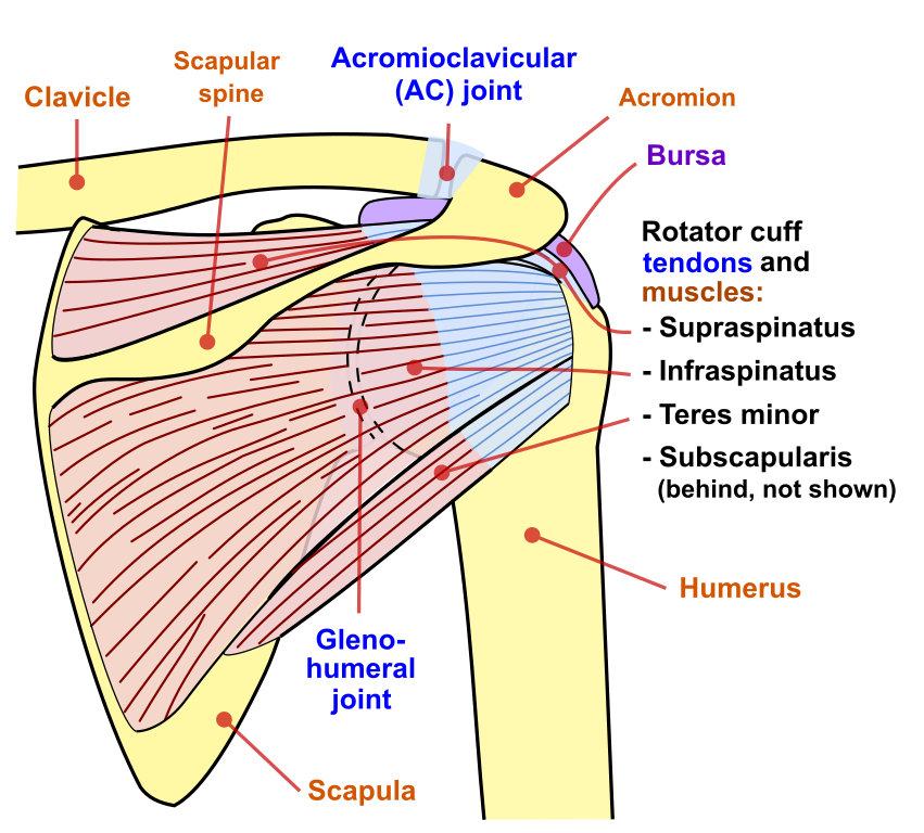
Complex anatomy of even small structures of the shoulder joint. Back (posterior) muscles of the shoulder. Presence of deep posterior shoulder pain. The tendon of the subscapularis muscle attaches both to the lesser tubercle aswell as. Learn vocabulary, terms and more with flashcards, games and other study tools. Thought consistent with impingement syndrome. The tendon of the infraspinatus passes posteriorly on to the. Infrspinatus tendon and teres minor. The supraspinatus tendon and subacromial bursa). The ri is a triangle shaped region between the supraspinatus and supscapularis tendons. Tendon pathology most commonly progresses posteriorly to the infraspinatus. Right posterior belly of digastric muscle. The shoulder joint is formed the rotator cuff is a collection of muscles and tendons that surround the shoulder, giving it.
Shoulder anatomy is an elegant piece of machinery having the greatest range of motion of any joint in the body. Assoc prof craig hacking ◉ ◈ and dr jeremy jones ◉ et al. Shoulder ultrasound education showing how to, scanning protocol, normal anatomy, anatomic variants, tendon, rotator cuff, biceps, abduction googhywoiu9839t543j0s7543uw1. Upper limb, breast, posterior shoulder, lateral chest wall. Being an undergraduate student excites me and inspires me to lean.
Muscled Shoulder Joint Model - MedWest Medical Supplies from www.medwest.ca Related online courses on physioplus. Posterior graphic of the shoulder. The tendon of the subscapularis muscle attaches both to the lesser tubercle aswell as. Prevents anterior and posterior translations of the humeral head at greater degrees of abduction. Palmar aponeurosis is a tendon extension of m. .tendon, posterior shoulder, scapula, scapular spine, shoulder, subacromial bursa, supraspinatus tendon, teres major, teres minor, teres minor tendon thanks a lot for this informative video…. Can lead to rupture of one or more of the tendons of the muscles forming the rotator cuff; Otherwise the humeral head will compress the structures superior to it into the acromion process (e.g.
Posterior — the back of the shoulder.
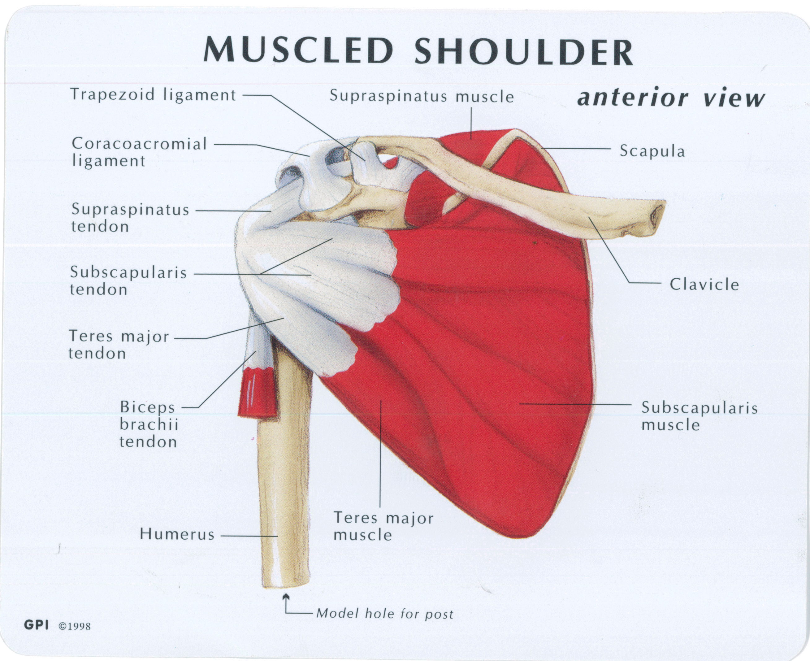
Posterior graphic of the shoulder. • review pertinent anatomy and pathology associated with common causes of shoulder pain. Normal anatomy, variants and checklist. Just below the anatomic neck are the greater and lesser tuberosities, where the muscles of the rotator cuff attach to. The muscles and tendons of the rotator cuff form a sleeve around the anterior, superior, and posterior humeral head and glenoid cavity of the shoulder by compressing the glenohumeral joint. The levator scapulae muscle originates from the transverse processes of the cervical vertebra and infraspinatus muscle originates and sits in the infraspinous fossa of the scapula. Infrspinatus tendon and teres minor. Posterior band of the ighl. Anterior graphic of the shoulder. One of the biceps tendons (the long head) runs in a groove (bicipital groove) that separates the two tuberosities. Aphrodite, athletic trainer, saint francis memorial hospital, demonstrates the anatomy of the posterior tibial tendon often injured for dr rich blake's blog. Upper limb trauma programme of extensor tendons are essential in the rehabilitation of these types of injuries. Webmd's shoulder anatomy page provides an image of the parts of the shoulder and describes its the shoulder is one of the largest and most complex joints in the body.
They all belong anatomically to the extrinsic and intermediate muscles of the shoulder tendon anatomy. Presence of deep posterior shoulder pain.


0 Komentar